

Which Of The Following Organelles Are Found In Both Animal And Plant Cells
Chapter 3: Introduction to Cell Structure and Part
iii.3 Eukaryotic Cells
By the end of this section, yous will be able to:
- Draw the construction of eukaryotic plant and animate being cells
- State the role of the plasma membrane
- Summarize the functions of the major cell organelles
- Depict the cytoskeleton and extracellular matrix
Sentinel a video nearly oxygen in the atmosphere.
At this betoken, it should be clear that eukaryotic cells accept a more than circuitous structure than exercise prokaryotic cells. Organelles allow for various functions to occur in the prison cell at the aforementioned time. Before discussing the functions of organelles within a eukaryotic jail cell, let us kickoff examine ii of import components of the prison cell: the plasma membrane and the cytoplasm.
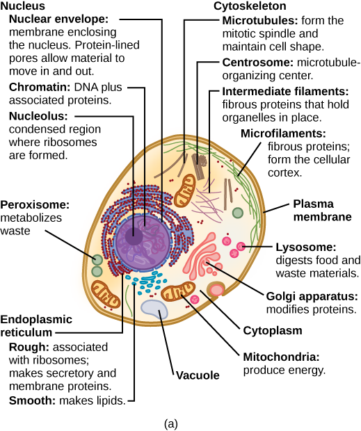
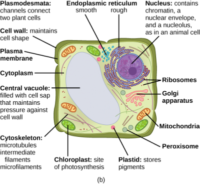
What structures does a plant prison cell have that an animate being cell does not take? What structures does an animal cell take that a plant cell does not take? Plant cells have plasmodesmata, a cell wall, a large cardinal vacuole, chloroplasts, and plastids. Beast cells accept lysosomes and centrosomes.
The Plasma Membrane
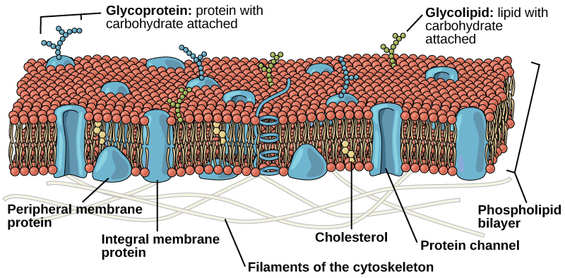
Like prokaryotes, eukaryotic cells have a plasma membrane (Effigy three.9) fabricated up of a phospholipid bilayer with embedded proteins that separates the internal contents of the cell from its surrounding environs. A phospholipid is a lipid molecule composed of ii fatty acid chains, a glycerol courage, and a phosphate group. The plasma membrane regulates the passage of some substances, such as organic molecules, ions, and h2o, preventing the passage of some to maintain internal weather condition, while actively bringing in or removing others. Other compounds move passively beyond the membrane.
The plasma membranes of cells that specialize in absorption are folded into fingerlike projections chosen microvilli (singular = microvillus). This folding increases the surface area of the plasma membrane. Such cells are typically found lining the small intestine, the organ that absorbs nutrients from digested food. This is an excellent example of form matching the function of a construction.
People with celiac affliction have an immune response to gluten, which is a protein establish in wheat, barley, and rye. The immune response damages microvilli, and thus, afflicted individuals cannot absorb nutrients. This leads to malnutrition, cramping, and diarrhea. Patients suffering from celiac illness must follow a gluten-gratuitous diet.
The Cytoplasm
The cytoplasm comprises the contents of a prison cell between the plasma membrane and the nuclear envelope (a structure to be discussed shortly). It is made up of organelles suspended in the gel-similar cytosol, the cytoskeleton, and diverse chemicals. Even though the cytoplasm consists of 70 to 80 percent h2o, it has a semi-solid consistency, which comes from the proteins inside it. Withal, proteins are not the only organic molecules institute in the cytoplasm. Glucose and other simple sugars, polysaccharides, amino acids, nucleic acids, fatty acids, and derivatives of glycerol are found in that location besides. Ions of sodium, potassium, calcium, and many other elements are also dissolved in the cytoplasm. Many metabolic reactions, including poly peptide synthesis, have place in the cytoplasm.
The Cytoskeleton
If you were to remove all the organelles from a cell, would the plasma membrane and the cytoplasm be the only components left? No. Within the cytoplasm, there would still be ions and organic molecules, plus a network of poly peptide fibers that helps to maintain the shape of the cell, secures certain organelles in specific positions, allows cytoplasm and vesicles to move within the cell, and enables unicellular organisms to move independently. Collectively, this network of protein fibers is known as the cytoskeleton. There are three types of fibers within the cytoskeleton: microfilaments, also known as actin filaments, intermediate filaments, and microtubules (Effigy 3.10).
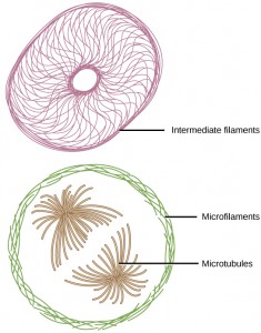
Microfilaments are the thinnest of the cytoskeletal fibers and function in moving cellular components, for example, during prison cell division. They as well maintain the structure of microvilli, the extensive folding of the plasma membrane establish in cells defended to absorption. These components are also common in muscle cells and are responsible for musculus cell contraction. Intermediate filaments are of intermediate diameter and have structural functions, such as maintaining the shape of the jail cell and anchoring organelles. Keratin, the compound that strengthens hair and nails, forms one blazon of intermediate filament. Microtubules are the thickest of the cytoskeletal fibers. These are hollow tubes that can dissolve and reform quickly. Microtubules guide organelle movement and are the structures that pull chromosomes to their poles during cell partitioning. They are also the structural components of flagella and cilia. In cilia and flagella, the microtubules are organized as a circumvolve of 9 double microtubules on the outside and ii microtubules in the eye.
The centrosome is a region near the nucleus of animal cells that functions every bit a microtubule-organizing center. It contains a pair of centrioles, two structures that lie perpendicular to each other. Each centriole is a cylinder of 9 triplets of microtubules.
The centrosome replicates itself before a cell divides, and the centrioles play a role in pulling the duplicated chromosomes to opposite ends of the dividing prison cell. Nonetheless, the exact office of the centrioles in cell sectionalization is not articulate, since cells that have the centrioles removed can still divide, and institute cells, which lack centrioles, are capable of cell sectionalization.
Flagella and Cilia
Flagella (atypical = flagellum) are long, hair-like structures that extend from the plasma membrane and are used to move an unabridged cell, (for case, sperm, Euglena). When present, the cell has just ane flagellum or a few flagella. When cilia (singular = cilium) are present, however, they are many in number and extend along the unabridged surface of the plasma membrane. They are brusk, hair-like structures that are used to move entire cells (such every bit paramecium) or motion substances along the outer surface of the cell (for example, the cilia of cells lining the fallopian tubes that move the ovum toward the uterus, or cilia lining the cells of the respiratory tract that move particulate matter toward the throat that mucus has trapped).
The Endomembrane System
The endomembrane system (endo = inside) is a group of membranes and organelles in eukaryotic cells that work together to modify, package, and transport lipids and proteins. Information technology includes the nuclear envelope, lysosomes, vesicles, endoplasmic reticulum and the Golgi apparatus, which we will cover shortly. Although not technically inside the cell, the plasma membrane is included in the endomembrane organization considering, as you will encounter, information technology interacts with the other endomembranous organelles.
The Nucleus
Typically, the nucleus is the most prominent organelle in a cell. The nucleus (plural = nuclei) houses the jail cell's DNA in the form of chromatin and directs the synthesis of ribosomes and proteins. Let u.s. look at it in more detail (Effigy iii.11).
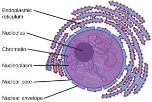
The nuclear envelope is a double-membrane structure that constitutes the outermost portion of the nucleus (Figure 3.xi). Both the inner and outer membranes of the nuclear envelope are phospholipid bilayers.
The nuclear envelope is punctuated with pores that control the passage of ions, molecules, and RNA betwixt the nucleoplasm and the cytoplasm.
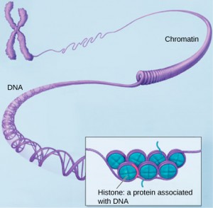
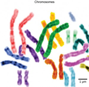
To empathise chromatin, information technology is helpful to showtime consider chromosomes. Chromosomes are structures within the nucleus that are made upward of DNA, the hereditary fabric, and proteins. This combination of Dna and proteins is called chromatin. In eukaryotes, chromosomes are linear structures. Every species has a specific number of chromosomes in the nucleus of its torso cells. For example, in humans, the chromosome number is 46, whereas in fruit flies, the chromosome number is 8.
Chromosomes are simply visible and distinguishable from one another when the prison cell is getting ready to divide. When the cell is in the growth and maintenance phases of its life cycle, the chromosomes resemble an unwound, jumbled bunch of threads.
We already know that the nucleus directs the synthesis of ribosomes, but how does it do this? Some chromosomes have sections of DNA that encode ribosomal RNA. A darkly stained expanse inside the nucleus, called the nucleolus (plural = nucleoli), aggregates the ribosomal RNA with associated proteins to gather the ribosomal subunits that are then transported through the nuclear pores into the cytoplasm.
The Endoplasmic Reticulum
The endoplasmic reticulum (ER) is a series of interconnected bleary tubules that collectively alter proteins and synthesize lipids. However, these ii functions are performed in separate areas of the endoplasmic reticulum: the rough endoplasmic reticulum and the shine endoplasmic reticulum, respectively.
The hollow portion of the ER tubules is called the lumen or cisternal space. The membrane of the ER, which is a phospholipid bilayer embedded with proteins, is continuous with the nuclear envelope.
The rough endoplasmic reticulum (RER) is so named considering the ribosomes fastened to its cytoplasmic surface give it a studded appearance when viewed through an electron microscope.
The ribosomes synthesize proteins while fastened to the ER, resulting in the transfer of their newly synthesized proteins into the lumen of the RER where they undergo modifications such as folding or addition of sugars. The RER too makes phospholipids for cell membranes.
If the phospholipids or modified proteins are not destined to stay in the RER, they will be packaged inside vesicles and transported from the RER by budding from the membrane. Since the RER is engaged in modifying proteins that volition be secreted from the cell, it is abundant in cells that secrete proteins, such equally the liver.
The smooth endoplasmic reticulum (SER) is continuous with the RER merely has few or no ribosomes on its cytoplasmic surface. The SER's functions include synthesis of carbohydrates, lipids (including phospholipids), and steroid hormones; detoxification of medications and poisons; booze metabolism; and storage of calcium ions.
The Golgi Apparatus
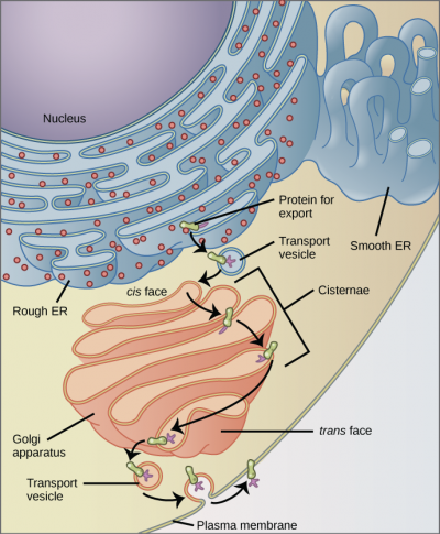
We accept already mentioned that vesicles can bud from the ER, simply where exercise the vesicles go? Before reaching their final destination, the lipids or proteins within the transport vesicles need to be sorted, packaged, and tagged so that they air current upwards in the right place. The sorting, tagging, packaging, and distribution of lipids and proteins take place in the Golgi appliance (also chosen the Golgi torso), a series of flattened membranous sacs.
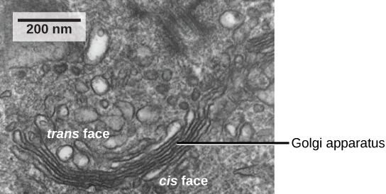
The Golgi appliance has a receiving face most the endoplasmic reticulum and a releasing face on the side away from the ER, toward the prison cell membrane. The ship vesicles that course from the ER travel to the receiving face, fuse with it, and empty their contents into the lumen of the Golgi apparatus. As the proteins and lipids travel through the Golgi, they undergo further modifications. The most frequent modification is the addition of short chains of sugar molecules. The newly modified proteins and lipids are then tagged with small-scale molecular groups to enable them to be routed to their proper destinations.
Finally, the modified and tagged proteins are packaged into vesicles that bud from the opposite face of the Golgi. While some of these vesicles, transport vesicles, deposit their contents into other parts of the cell where they will exist used, others, secretory vesicles, fuse with the plasma membrane and release their contents exterior the cell.
The amount of Golgi in different cell types over again illustrates that course follows function inside cells. Cells that engage in a great deal of secretory activeness (such equally cells of the salivary glands that secrete digestive enzymes or cells of the immune system that secrete antibodies) accept an abundant number of Golgi.
In establish cells, the Golgi has an additional role of synthesizing polysaccharides, some of which are incorporated into the prison cell wall and some of which are used in other parts of the cell.
Lysosomes
In animal cells, the lysosomes are the jail cell'due south "garbage disposal." Digestive enzymes within the lysosomes aid the breakdown of proteins, polysaccharides, lipids, nucleic acids, and even worn-out organelles. In single-celled eukaryotes, lysosomes are important for digestion of the food they ingest and the recycling of organelles. These enzymes are active at a much lower pH (more acidic) than those located in the cytoplasm. Many reactions that have place in the cytoplasm could not occur at a depression pH, thus the advantage of compartmentalizing the eukaryotic prison cell into organelles is credible.
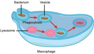
Lysosomes also employ their hydrolytic enzymes to destroy disease-causing organisms that might enter the cell. A good example of this occurs in a grouping of white claret cells chosen macrophages, which are office of your body's immune system. In a process known as phagocytosis, a section of the plasma membrane of the macrophage invaginates (folds in) and engulfs a pathogen. The invaginated section, with the pathogen within, then pinches itself off from the plasma membrane and becomes a vesicle. The vesicle fuses with a lysosome. The lysosome's hydrolytic enzymes and then destroy the pathogen (Figure 3.xv).
Vesicles and Vacuoles
Vesicles and vacuoles are membrane-spring sacs that function in storage and transport. Vacuoles are somewhat larger than vesicles, and the membrane of a vacuole does not fuse with the membranes of other cellular components. Vesicles tin can fuse with other membranes within the cell organisation. Additionally, enzymes within plant vacuoles tin can suspension downwards macromolecules.
Why does the cis confront of the Golgi non face the plasma membrane?
<!– Considering that face receives chemicals from the ER, which is toward the center of the cell. –>
Ribosomes
Ribosomes are the cellular structures responsible for protein synthesis. When viewed through an electron microscope, gratis ribosomes appear as either clusters or single tiny dots floating freely in the cytoplasm. Ribosomes may be fastened to either the cytoplasmic side of the plasma membrane or the cytoplasmic side of the endoplasmic reticulum. Electron microscopy has shown that ribosomes consist of big and small-scale subunits. Ribosomes are enzyme complexes that are responsible for protein synthesis.
Because poly peptide synthesis is essential for all cells, ribosomes are constitute in practically every cell, although they are smaller in prokaryotic cells. They are especially abundant in immature blood-red blood cells for the synthesis of hemoglobin, which functions in the send of oxygen throughout the body.
Mitochondria
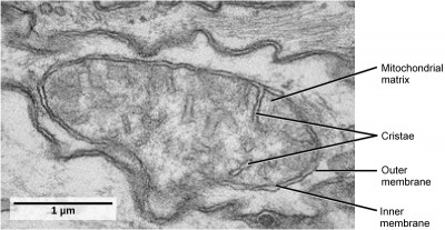
Mitochondria (singular = mitochondrion) are often called the "powerhouses" or "free energy factories" of a cell because they are responsible for making adenosine triphosphate (ATP), the prison cell's master free energy-carrying molecule. The formation of ATP from the breakup of glucose is known equally cellular respiration. Mitochondria are oval-shaped, double-membrane organelles (Figure 3.17) that take their own ribosomes and Deoxyribonucleic acid. Each membrane is a phospholipid bilayer embedded with proteins. The inner layer has folds called cristae, which increment the surface area of the inner membrane. The area surrounded past the folds is called the mitochondrial matrix. The cristae and the matrix have different roles in cellular respiration.
In keeping with our theme of form post-obit function, information technology is important to point out that muscle cells have a very high concentration of mitochondria because muscle cells need a lot of energy to contract.
Peroxisomes
Peroxisomes are pocket-size, round organelles enclosed by single membranes. They carry out oxidation reactions that break down fat acids and amino acids. They as well detoxify many poisons that may enter the torso. Alcohol is detoxified past peroxisomes in liver cells. A byproduct of these oxidation reactions is hydrogen peroxide, H2O2, which is contained inside the peroxisomes to prevent the chemical from causing impairment to cellular components outside of the organelle. Hydrogen peroxide is safely broken down past peroxisomal enzymes into h2o and oxygen.
Beast Cells versus Found Cells
Despite their fundamental similarities, there are some striking differences between animal and plant cells (come across Table iii.1). Fauna cells accept centrioles, centrosomes (discussed under the cytoskeleton), and lysosomes, whereas plant cells practice not. Plant cells have a cell wall, chloroplasts, plasmodesmata, and plastids used for storage, and a large fundamental vacuole, whereas animal cells do not.
The Jail cell Wall
In Figure 3.eightb, the diagram of a plant prison cell, yous see a structure external to the plasma membrane called the prison cell wall. The cell wall is a rigid covering that protects the cell, provides structural support, and gives shape to the cell. Fungal and protist cells also have cell walls.
While the primary component of prokaryotic cell walls is peptidoglycan, the major organic molecule in the plant cell wall is cellulose, a polysaccharide made up of long, straight bondage of glucose units. When nutritional information refers to dietary cobweb, it is referring to the cellulose content of food.
Chloroplasts
Like mitochondria, chloroplasts too accept their ain Deoxyribonucleic acid and ribosomes. Chloroplasts function in photosynthesis and can be institute in eukaryotic cells such equally plants and algae. In photosynthesis, carbon dioxide, water, and light energy are used to brand glucose and oxygen. This is the major departure between plants and animals: Plants (autotrophs) are able to brand their own food, like glucose, whereas animals (heterotrophs) must rely on other organisms for their organic compounds or food source.
Like mitochondria, chloroplasts have outer and inner membranes, only within the space enclosed by a chloroplast's inner membrane is a ready of interconnected and stacked, fluid-filled membrane sacs called thylakoids (Figure 3.eighteen). Each stack of thylakoids is called a granum (plural = grana). The fluid enclosed by the inner membrane and surrounding the grana is called the stroma.
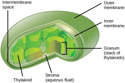
The chloroplasts contain a green pigment called chlorophyll, which captures the energy of sunlight for photosynthesis. Similar constitute cells, photosynthetic protists also have chloroplasts. Some leaner also perform photosynthesis, just they do non take chloroplasts. Their photosynthetic pigments are located in the thylakoid membrane inside the cell itself.
Evolution in Activeness
Endosymbiosis: We have mentioned that both mitochondria and chloroplasts contain DNA and ribosomes. Have you wondered why? Stiff evidence points to endosymbiosis equally the caption.
Symbiosis is a relationship in which organisms from two separate species alive in close clan and typically exhibit specific adaptations to each other. Endosymbiosis (endo-= inside) is a relationship in which i organism lives inside the other. Endosymbiotic relationships grow in nature. Microbes that produce vitamin K alive within the human gut. This human relationship is beneficial for us because we are unable to synthesize vitamin Yard. It is also beneficial for the microbes because they are protected from other organisms and are provided a stable habitat and abundant food by living within the large intestine.
Scientists take long noticed that bacteria, mitochondria, and chloroplasts are similar in size. We also know that mitochondria and chloroplasts have Deoxyribonucleic acid and ribosomes, but as bacteria do and they resemble the types found in bacteria. Scientists believe that host cells and bacteria formed a mutually beneficial endosymbiotic relationship when the host cells ingested aerobic leaner and blue-green alga but did not destroy them. Through development, these ingested bacteria became more specialized in their functions, with the aerobic bacteria becoming mitochondria and the photosynthetic bacteria becoming chloroplasts.
The Fundamental Vacuole
Previously, we mentioned vacuoles as essential components of plant cells. If you wait at Figure three.8b, you volition see that found cells each take a large, central vacuole that occupies about of the cell. The central vacuole plays a key role in regulating the cell's concentration of water in irresolute ecology conditions. In plant cells, the liquid inside the fundamental vacuole provides turgor pressure level, which is the outward pressure caused past the fluid inside the cell. Take you always noticed that if you lot forget to water a found for a few days, it wilts? That is because as the h2o concentration in the soil becomes lower than the water concentration in the plant, water moves out of the central vacuoles and cytoplasm and into the soil. As the central vacuole shrinks, information technology leaves the cell wall unsupported. This loss of support to the jail cell walls of a found results in the wilted appearance. Additionally, this fluid has a very biting gustatory modality, which discourages consumption by insects and animals. The central vacuole also functions to store proteins in developing seed cells.
Extracellular Matrix of Animal Cells
Nigh animate being cells release materials into the extracellular infinite. The primary components of these materials are glycoproteins and the protein collagen. Collectively, these materials are chosen the extracellular matrix (Figure 3.19). Not simply does the extracellular matrix hold the cells together to form a tissue, but it as well allows the cells within the tissue to communicate with each other.
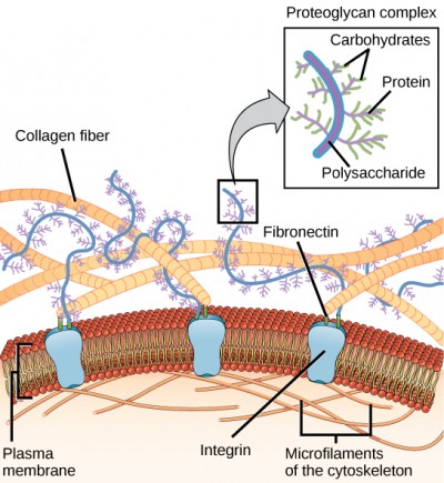
Blood clotting provides an example of the role of the extracellular matrix in jail cell advice. When the cells lining a blood vessel are damaged, they brandish a protein receptor called tissue cistron. When tissue factor binds with another factor in the extracellular matrix, it causes platelets to adhere to the wall of the damaged blood vessel, stimulates next shine muscle cells in the blood vessel to contract (thus constricting the claret vessel), and initiates a series of steps that stimulate the platelets to produce clotting factors.
Intercellular Junctions
Cells can besides communicate with each other by direct contact, referred to as intercellular junctions. At that place are some differences in the means that establish and brute cells do this. Plasmodesmata (singular = plasmodesma) are junctions betwixt establish cells, whereas animate being cell contacts include tight and gap junctions, and desmosomes.
In general, long stretches of the plasma membranes of neighboring plant cells cannot affect 1 some other because they are separated by the prison cell walls surrounding each cell. Plasmodesmata are numerous channels that laissez passer between the cell walls of adjacent plant cells, connecting their cytoplasm and enabling signal molecules and nutrients to be transported from jail cell to jail cell (Figure 3.20a).
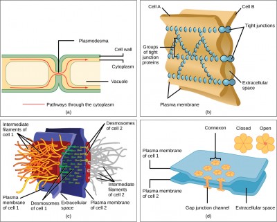
A tight junction is a watertight seal between two adjacent animal cells (Figure 3.twentyb). Proteins concur the cells tightly against each other. This tight adhesion prevents materials from leaking betwixt the cells. Tight junctions are typically found in the epithelial tissue that lines internal organs and cavities, and composes near of the peel. For case, the tight junctions of the epithelial cells lining the urinary bladder preclude urine from leaking into the extracellular infinite.
Also found only in beast cells are desmosomes, which act like spot welds between adjacent epithelial cells (Effigy 3.20c). They keep cells together in a sheet-like formation in organs and tissues that stretch, like the skin, middle, and muscles.
Gap junctions in animal cells are like plasmodesmata in plant cells in that they are channels betwixt adjacent cells that allow for the transport of ions, nutrients, and other substances that enable cells to communicate (Effigy 3.20d). Structurally, however, gap junctions and plasmodesmata differ.
Cell Component | Office | Present in Prokaryotes? | Present in Animal Cells? | Present in Constitute Cells? |
|---|---|---|---|---|
| Plasma membrane | Separates prison cell from external environment; controls passage of organic molecules, ions, water, oxygen, and wastes into and out of the prison cell | Yep | Yeah | Aye |
| Cytoplasm | Provides structure to jail cell; site of many metabolic reactions; medium in which organelles are found | Yes | Yes | Yes |
| Nucleoid | Location of Dna | Yes | No | No |
| Nucleus | Jail cell organelle that houses Deoxyribonucleic acid and directs synthesis of ribosomes and proteins | No | Yes | Yes |
| Ribosomes | Protein synthesis | Yes | Yeah | Yes |
| Mitochondria | ATP production/cellular respiration | No | Yes | Yes |
| Peroxisomes | Oxidizes and breaks down fatty acids and amino acids, and detoxifies poisons | No | Aye | Yep |
| Vesicles and vacuoles | Storage and transport; digestive function in plant cells | No | Yes | Yes |
| Centrosome | Unspecified function in jail cell sectionalisation in beast cells; organizing center of microtubules in animal cells | No | Yes | No |
| Lysosomes | Digestion of macromolecules; recycling of worn-out organelles | No | Yes | No |
| Cell wall | Protection, structural support and maintenance of prison cell shape | Yep, primarily peptidoglycan in bacteria merely not Archaea | No | Yeah, primarily cellulose |
| Chloroplasts | Photosynthesis | No | No | Aye |
| Endoplasmic reticulum | Modifies proteins and synthesizes lipids | No | Yeah | Yes |
| Golgi appliance | Modifies, sorts, tags, packages, and distributes lipids and proteins | No | Yes | Yes |
| Cytoskeleton | Maintains cell'southward shape, secures organelles in specific positions, allows cytoplasm and vesicles to move within the cell, and enables unicellular organisms to move independently | Yes | Yes | Aye |
| Flagella | Cellular locomotion | Some | Some | No, except for some plant sperm. |
| Cilia | Cellular locomotion, motion of particles forth extracellular surface of plasma membrane, and filtration | No | Some | No |
Section Summary
Like a prokaryotic cell, a eukaryotic jail cell has a plasma membrane, cytoplasm, and ribosomes, but a eukaryotic cell is typically larger than a prokaryotic cell, has a true nucleus (pregnant its DNA is surrounded by a membrane), and has other membrane-bound organelles that allow for compartmentalization of functions. The plasma membrane is a phospholipid bilayer embedded with proteins. The nucleolus within the nucleus is the site for ribosome assembly. Ribosomes are found in the cytoplasm or are fastened to the cytoplasmic side of the plasma membrane or endoplasmic reticulum. They perform protein synthesis. Mitochondria perform cellular respiration and produce ATP. Peroxisomes suspension down fatty acids, amino acids, and some toxins. Vesicles and vacuoles are storage and transport compartments. In plant cells, vacuoles also help suspension down macromolecules.
Animal cells also have a centrosome and lysosomes. The centrosome has two bodies, the centrioles, with an unknown role in prison cell division. Lysosomes are the digestive organelles of animal cells.
Plant cells have a cell wall, chloroplasts, and a central vacuole. The plant cell wall, whose primary component is cellulose, protects the prison cell, provides structural support, and gives shape to the prison cell. Photosynthesis takes identify in chloroplasts. The central vacuole expands, enlarging the cell without the demand to produce more cytoplasm.
The endomembrane system includes the nuclear envelope, the endoplasmic reticulum, Golgi appliance, lysosomes, vesicles, also as the plasma membrane. These cellular components work together to modify, packet, tag, and ship membrane lipids and proteins.
The cytoskeleton has three different types of protein elements. Microfilaments provide rigidity and shape to the cell, and facilitate cellular movements. Intermediate filaments bear tension and ballast the nucleus and other organelles in identify. Microtubules aid the jail cell resist compression, serve as tracks for motor proteins that move vesicles through the cell, and pull replicated chromosomes to opposite ends of a dividing cell. They are also the structural elements of centrioles, flagella, and cilia.
Fauna cells communicate through their extracellular matrices and are connected to each other past tight junctions, desmosomes, and gap junctions. Plant cells are continued and communicate with each other by plasmodesmata.
prison cell wall: a rigid cell covering fabricated of cellulose in plants, peptidoglycan in bacteria, not-peptidoglycan compounds in Archaea, and chitin in fungi that protects the cell, provides structural support, and gives shape to the cell
central vacuole: a big plant cell organelle that acts every bit a storage compartment, water reservoir, and site of macromolecule deposition
chloroplast: a plant cell organelle that carries out photosynthesis
cilium: (plural: cilia) a brusk, pilus-like structure that extends from the plasma membrane in big numbers and is used to motility an entire cell or move substances along the outer surface of the jail cell
cytoplasm: the entire region between the plasma membrane and the nuclear envelope, consisting of organelles suspended in the gel-like cytosol, the cytoskeleton, and various chemicals
cytoskeleton: the network of protein fibers that collectively maintains the shape of the prison cell, secures some organelles in specific positions, allows cytoplasm and vesicles to move within the cell, and enables unicellular organisms to move
cytosol: the gel-like material of the cytoplasm in which cell structures are suspended
desmosome: a linkage between side by side epithelial cells that forms when cadherins in the plasma membrane attach to intermediate filaments
endomembrane organization: the group of organelles and membranes in eukaryotic cells that work together to change, packet, and ship lipids and proteins
endoplasmic reticulum (ER): a series of interconnected membranous structures inside eukaryotic cells that collectively modify proteins and synthesize lipids
extracellular matrix: the fabric, primarily collagen, glycoproteins, and proteoglycans, secreted from beast cells that holds cells together as a tissue, allows cells to communicate with each other, and provides mechanical protection and anchoring for cells in the tissue
flagellum: (plural: flagella) the long, hair-like structure that extends from the plasma membrane and is used to motility the jail cell
gap junction: a channel between ii next animal cells that allows ions, nutrients, and other low-molecular weight substances to pass between the cells, enabling the cells to communicate
Golgi apparatus: a eukaryotic organelle made upwards of a series of stacked membranes that sorts, tags, and packages lipids and proteins for distribution
lysosome: an organelle in an creature cell that functions as the cell's digestive component; it breaks downwardly proteins, polysaccharides, lipids, nucleic acids, and even worn-out organelles
mitochondria: (singular: mitochondrion) the cellular organelles responsible for carrying out cellular respiration, resulting in the production of ATP, the cell's main energy-conveying molecule
nuclear envelope: the double-membrane structure that constitutes the outermost portion of the nucleus
nucleolus: the darkly staining torso inside the nucleus that is responsible for assembling ribosomal subunits
nucleus: the cell organelle that houses the cell's DNA and directs the synthesis of ribosomes and proteins
peroxisome: a pocket-sized, round organelle that contains hydrogen peroxide, oxidizes fatty acids and amino acids, and detoxifies many poisons
plasma membrane: a phospholipid bilayer with embedded (integral) or attached (peripheral) proteins that separates the internal contents of the cell from its surrounding environment
plasmodesma: (plural: plasmodesmata) a channel that passes between the cell walls of adjacent plant cells, connects their cytoplasm, and allows materials to be transported from cell to cell
ribosome: a cellular structure that carries out protein synthesis
rough endoplasmic reticulum (RER): the region of the endoplasmic reticulum that is studded with ribosomes and engages in protein modification
smooth endoplasmic reticulum (SER): the region of the endoplasmic reticulum that has few or no ribosomes on its cytoplasmic surface and synthesizes carbohydrates, lipids, and steroid hormones; detoxifies chemicals like pesticides, preservatives, medications, and environmental pollutants, and stores calcium ions
tight junction: a firm seal between two adjacent brute cells created by poly peptide adherence
vacuole: a membrane-jump sac, somewhat larger than a vesicle, that functions in cellular storage and transport
vesicle: a small, membrane-bound sac that functions in cellular storage and transport; its membrane is capable of fusing with the plasma membrane and the membranes of the endoplasmic reticulum and Golgi apparatus
Media Attribution
- Effigy three.11: modification of work by NIGMS, NIH
- Effigy 3.13: modification of work by NIH; scale-bar information from Matt Russell
- Figure 3.fourteen: modification of piece of work past Louisa Howard; scale-bar data from Matt Russell
- Effigy 3.sixteen: modification of piece of work by Magnus Manske
- Figure 3.17: modification of piece of work past Matthew Britton; scale-bar data from Matt Russell
- Figure 3.twenty: modification of work by Mariana Ruiz Villareal
Source: https://opentextbc.ca/biology/chapter/3-3-eukaryotic-cells/
Posted by: thomasthadvating.blogspot.com
0 Response to "Which Of The Following Organelles Are Found In Both Animal And Plant Cells"
Post a Comment